Overview:
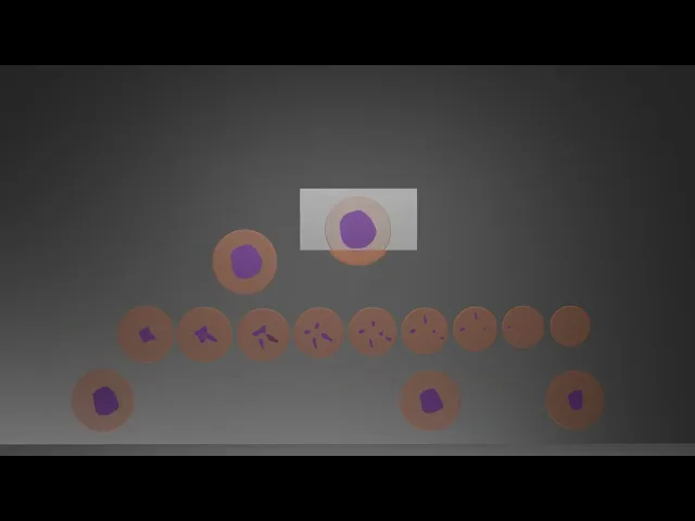
The videos illustrate how the surgically removed tumors, along with the small safety margin of healthy skin, are cut into wafer-thin slices, stained, and examined under the microscope. This example shows a tumor completely removed at first attempt. In the second example, the lateral incision edge, and in the third example the deep incision edge, both show residual tumor. Consequently, precise additional cutting must be performed at these sites.
To see the other two examples click here:
Use Case:
Used to explain the procedure in discussions with patients.
Used as information of the procedure, accessible on the Website of the University Hospital Zürich.
Creative Approach:
The focus was on clarity over aesthetics. I designed the animation with the patients in mind, aiming to communicate the surgical logic as simply and effectively as possible, so viewers can quickly understand how and why the procedure is performed.
Client/Collaboration:
Prof. Dr. med. Jürg Hafner
Universitätsspital Zürich